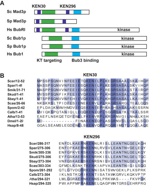
Figure 1. Mad3p contains two conserved KEN boxes.
A) Mad3/BubR1 domain structure–schematic diagrams of the Mad3 and Bub proteins indicate the organisation of their functional domains. B) Clustal×alignment of the two conserved KEN boxes found in Mad3/BubR1. Species indicated are Saccharomyces cerevisiae (Scer), Saccharomyces paradoxus (Spar), Saccharomyces mikatae (Smik), Saccharomyces kudriavzevii (Skud), Saccharomyces bayanus (Sbay), Saccharomyces castellii (Scas), Schizosaccharomyces pombe (Spom), Candida albicans (Calb), Arabidopsis thaliana (Atha), Drosophila melanogaster (Dmel) and Homo sapiens (Hsap). Numbers indicate residue position within protein sequence.

