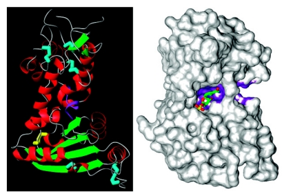
Figure 2.
Crystal Structures of CD38. Left panel. Color codes for the secondary structures are: green, β-structures; red, α-helix; cyan, conserved disulfide bonds; yellow, unique disulfide bond; purple, TLEDTL-conserved motif. Right panel. The van der Waal’s surface of CD38 is shown. The bound substrate molecule at the active site is nicotinamide mononucleotide. The surface of the TLEDTL-conserved motif is colored purple.

