
Figure 3.
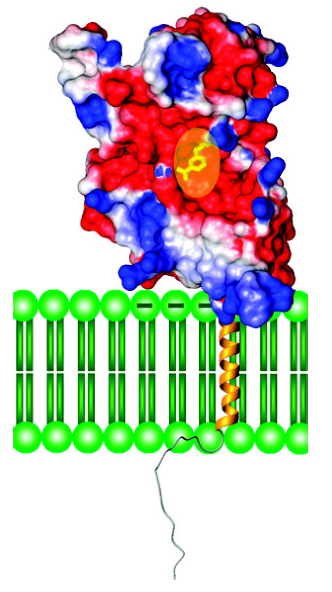
A model of native CD38 embedded in a membrane. The surface potential of the extra-membrane domain is based on crystallography, with the negative charged regions colored red, while positively charged are blue. The transparent oval (yellow) indicates the location of the active site pocket. The transmembrane segment is modeled arbitrarily as a helix (gold), while the N-terminal tail as a random coil. Phospholipids of the membrane are colored green.