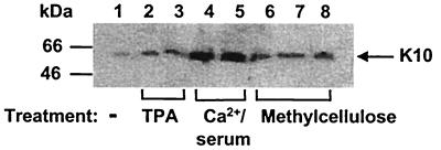
Figure 1.
Analysis of keratin 10 expression in W12 cells. Cells were untreated (lane 1), or treated with 16 nM PMA for 5 days (lane 2) and 10 days (lane 3), increased Ca2+ and serum concentration for 5 days (lane 4) or 10 days (lane 5), or suspended in methylcellulose for 1 day (lane 6), 2 days (lane 7), or 8 days (lane 8). Total extracts (50 μg) were used and Western blot analysis was performed with the anti-K10 Ck 8.60 antibody.

