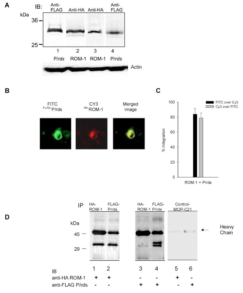
Fig. 1. Expression of FLAG-P/rds and HA-ROM-1 in Cos-7 cells.

A. levels of FLAG-P/rds and HA-ROM-1 expression in COS 7 cells. Levels of P/rds and ROM-1 protein expression were compared in cell extracts prepared as described in the methods from Lane1- cells transfected with only FLAG-P/rds, Lane 2, Cells transfected with only HA-ROM-1, Lane 3- cells co-transfected with both FLAG-P/rds and HA-ROM-1, probed with anti-HA antibody and Lane 4 co-transfected cells probed with anti-FLAG Ab. Similar levels of total protein are expressed in all three transfections. Bottom portion of figure, actin protein loading controls, membranes were stripped and re-probed with 1:250 dilution of anti-actin antibody (Santa Cruz Biotechnology). B. Co-localization of FLAG-P/rds and HA-ROM-1. COS cells were co-transfected with FLAG- P/rds and HA-ROM-1, permeabilized and immuno-stained. FLAG-P/rds was detected with M5 anti-FLAG and FITC conjugated goat-anti-mouse secondary antibody. HA-ROM-1 was detected using HA polyclonal antibody CY-3 conjugated secondary Ab. All images were captured with the same laser settings. C. Quantitation of co-localization. Analysis of fluorescent probe co-localization was performed using image analysis software [Metamorph; (Universal Imaging Corporation; Downingtown, PA), ver.6]. Regions of interest were defined to include cells that did not overlap. The region was segmented to select pixels above a constant threshold value (>60% above background) which represent true fluorescence. Since both spatial location and intensity of pixels contribute to co-localization, the values represent the integrated intensity; pixels in both Cy-3 and FITC images had similar brightness values and spatial location. The average pixel intensity for each is presented as either FIOTC over Cy-3 (black bars) or Cy-3 over FITC (grey bars). Co-localization analysis was performed on all cells present in Figure 1B. D. Co-immunoprecipitation of FLAG-P/rds and HA-ROM-1. COS-7 cells transfected with FLAG-P/rds and HA-ROM-1 were harvested, cell extracts prepared and immuno-precipitated with M5 monoclonal anti-FLAG-Ab or anti-HA antibody. Immunoprecipitates were fractionated and immuno-blotted with either monoclonal anti-HA Ab (lanes 1, 2 and 5) or monoclonal anti-FLAG Ab (lanes 3, 4 and 6) as indicated. Negative MOP-C21 controls are shown in lanes 5 and 6. Marker sizes in kDa are indicated on the left.
