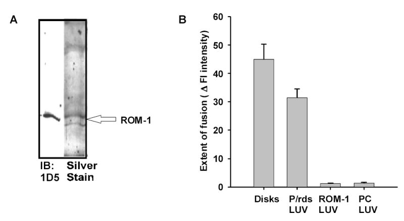
Fig. 3. Assessment of ROM-1 fusogenicity.

A. Isolation of ROM-1 from bovine OS membranes. ROM-1 was purified using a strategy identical to that described for P/RDS and described in detail in the Methods. Purified ROM-1 isolated in fractions 75 to 82 was run on SDS-Gels (10%) and silver stained (lane 1). An aliquot of the fraction (lane 2) was also run on SDS-PAGE and transferred to nitrocellulose and labeled with anti-ROM-1 antibody 1D5 (a generous gift from Dr. Robert Molday). B. Fusion between R18-PM and unlabeled target membranes; analysis of final extent of fusion. The extent of fusion between R18-PM and LUV containing either P/rds or ROM-1 was followed at 37oC. The extent of fusion is determined as the % change in fluorescence intensity over a 60 minute time period. During this period, fusion between R18-PM and disks goes to completion (Boesze-Battaglia, Albert et al. 1992; Boesze-Battaglia 1997). Results are the mean +/− SEM for three independent preparations each in duplicate
