
Fig. 4. A. Western blot of ROM-1-P/rds enriched vesicles.
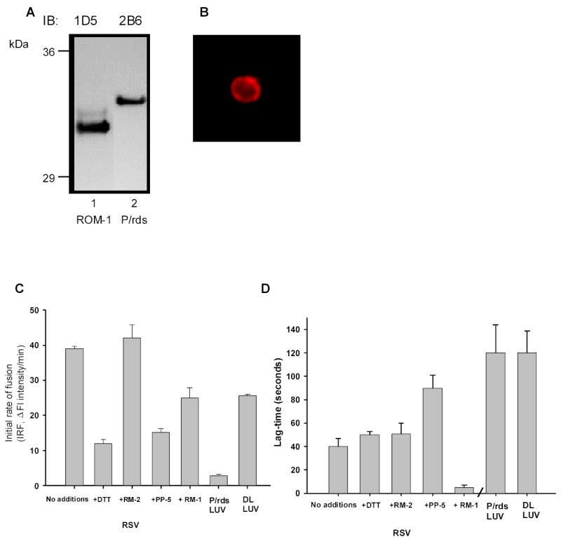
RSV were prepared as described in the Methods, separated by SDS- PAGE transferred and immunoblottted with either anit-P/RDS mAb 2B6 or anti-ROM-1 mAb 1D5. B. FITC labeling of ROM-1 P/rds Enriched Vesicles (RSV). Rim specific vesicles (RSV’s) were prepared from the dialyzed lipid rich fraction of ROS–disk Con A chromatography by freeze/thawing, labeled with anti-P/RDS mAb antibody 2B6 and imaged as described in the Methods. C. Fusion kinetics between R18-PM and unlabeled target membranes; analysis of IRF. Initial Rates of Fusion (IRF) are shown for fusion between R18-PM and disk membranes as well as RSV. RSV fusion is shown on the left. RSV –R18-PM fusion with no-additions RSV pre-incubated with 10 mM DTT, 10μg/ml PP-5 peptide or 10 μg/ml RM-1 or RM-2 peptide as indicated. Results are the mean +/− SEM for three independent preparations each in duplicate. Controls showing fusion between R18-PM and either P/rds containing LUV or DL LUV is indicated on the right in the last two columns. D. Fusion kinetics between R18-PM and unlabeled target membranes; analysis of lag-time. Lag-times are shown for fusion between R18-PM and RSV on the left. Lag-times observed when RSV fuse with R18-PM fusion with no-additions or RSV pre-incubated with 10 mM DTT, 10μg/ml PP-5 peptide or 10 μg/ml RM-1 or RM-2 peptide as indicated. Results are the mean +/− SEM for three independent preparations each in duplicate. Controls showing lag-time when fusion between R18-PM and either P/rds containing LUV or DL LUV is indicated on the right in the last two columns. Results are the mean +/− SEM for three independent preparations each in duplicate.