
Figure 1.
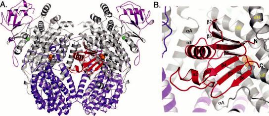
(A) Global structure of the PHH–PHM complex and (B) enlarged view of PHM in the PHH canyon. The PHH α-, β-, and γ-subunits are colored gray, purple, and pink, respectively, and PHM is colored red. Iron and zinc atoms are depicted as orange and green spheres, respectively. All figures were generated by using PyMOL (70).