
Figure 7.
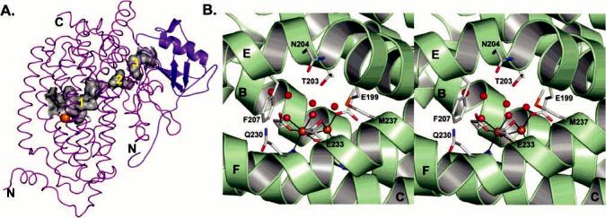
Cavities and pores in the PHH α-subunit. (A) α-subunit cavities (gray) that extend from the diiron center to the γ-subunit (blue). (B) Stereoview of the conserved residues and water molecules (red spheres) contributing to the pore through the α-subunit four-helix bundle.