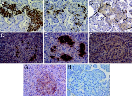
Fig. 1.
A type 1 diabetic pancreas shows nondestructive insulitis with NK cell infiltration and IFNα expression. (A–C) Immunohistochemistry panel showing reactivity for insulin (A), NK cells (B), and CD3+ cells (C) in consecutive sections representing the same islet; arrows point to scattered CD3+ cells (patient 1). (D–F) Reactivity to insulin in pancreatic sections from three additional type 1 diabetic patients (patients 4–6) showing a significant reduction of insulin-positive cells in these three individuals. Positivity for IFNα was detected in patient 1 (G) and in patients 2 and 3 (not shown), but not in control individuals (H). (Magnification: ×250.)

