
FIGURE 3.
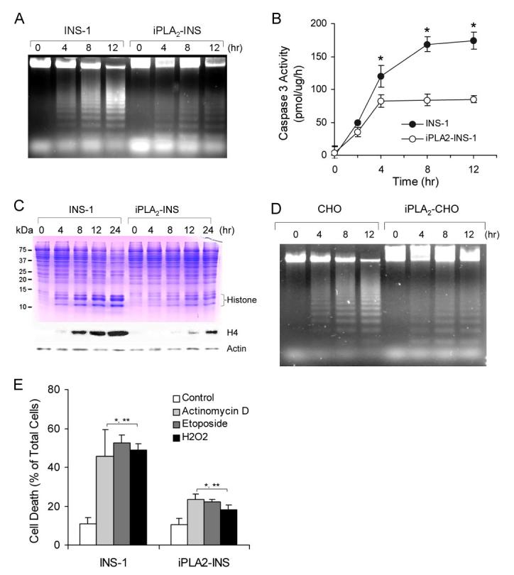
iPLA2 prevents apoptosis induced by mitochondrially generated superoxide. A, apoptotic DNA ladder analysis of INS-1 cells. After 1 μM STS treatment as in Fig. 1A, DNA was purified by an Apoptotic DNA ladder kit and analyzed on an agarose gel. B, comparison of caspase 3 activity between INS-1 and iPLA2-INS cells during STS-induced apoptosis. Caspase 3 activity was determined by a CaspACE assay system (n = 6). *, p < 0.05. C, accumulation of histones in the cytoplasm of the cells. After treatment with 1 μM STS for the indicated times, cytosol from both cell lines was prepared and analyzed by SDS-PAGE (upper) and Western blot for histone 4 (lower). D, apoptotic DNA ladder analysis in CHO cells. Mock-transfected CHO cells and CHO cells transfected with pcDNA3-iPLA2 were treated with 1 μM STS for the indicated times. The cells were collected, and DNA was prepared as in A and analyzed on an agarose gel. E, comparison of actinomycin D-, etoposide-, and H2O2-induced apoptosis between INS-1 and iPLA2-INS cells. Cells were treated with actinomycin D etoposide for 8 h and with H2O2 for 4 h. *, compared with control; **, compared between INS-1 and iPLA2-INS cells. p < 0.05.