
Figure 5.
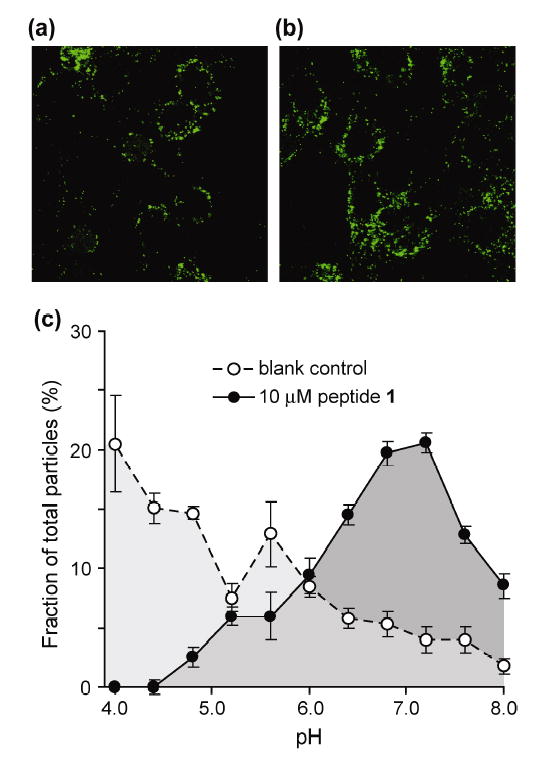
(a) Representative fluorescence microscopy image of cells infected with CF-Ad2-ts1; (b) representative image of cells infected with CF-Ad2-ts1 in the presence of 10 μM peptide 1; (c) population distribution of environmental pH for particles from images taken in the presence or absence of peptide 1.