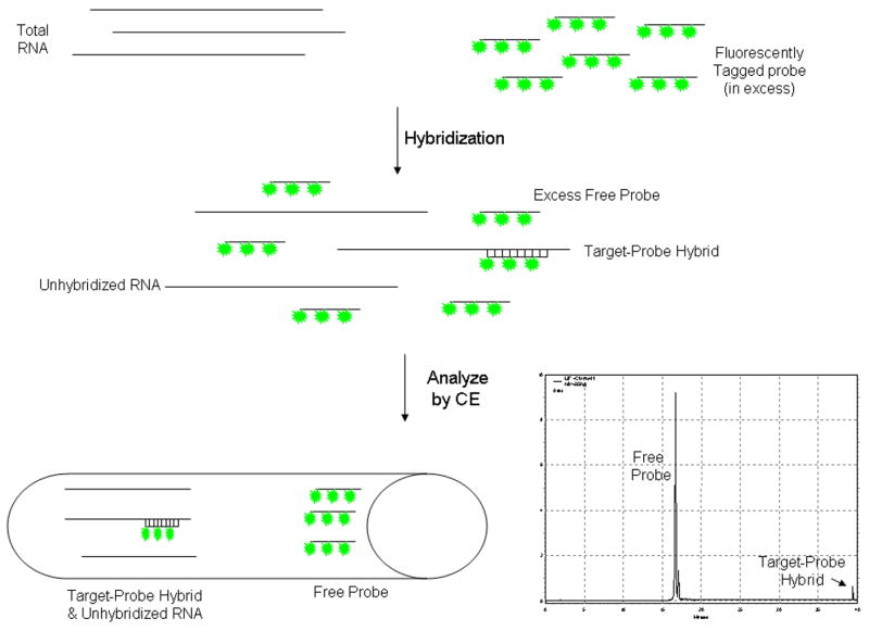
Figure 1.
Schematic Using CE-LIF to Quantify RNA Expression. Fluorescently tagged riboprobe is added in excess to a sample of RNA and allowed to undergo hybridization of the labeled riboprobe to target RNA. The sample, containing free probe, unhybridized RNA and probe-target hybrids, is injected into a silica capillary containing a sieving matrix which will separate the various components of the hybridization reaction based upon their size. Using a fluorescence detector, as the various components of the hybridization reaction pass through the detection window, only those with the fluorescent molecule incorporated (free probe or probe-target hybrids) will be detected. Since the probe is much smaller than the target-probe hybrid it passes through the detection window first followed at a later time by the target-probe hybrid.

