
Figure 3.
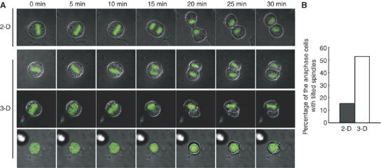
The cell–substrate adhesion regulates spindle orientation in the cells cultured in three dimensions. (A) Time-lapse images of the mitotic GFP-H2B-expressing HeLa cells that were cultured on top of (2-D) or within (3-D) a gel of a basement membrane matrix. The merge images of phase contrast and GFP-H2B are shown. For 3-D culture, three typical images of the cells are shown; the axes of the spindles were parallel (3-D, upper), tilted (3-D, middle), or perpendicular (3-D, bottom) to the horizontal plane. (B) Percentage of the cells that separate their chromosomes along the axis of their spindles which is tilted or perpendicular to the horizontal plane (2-D; n=72, 3-D; n=34).