
Figure 6.
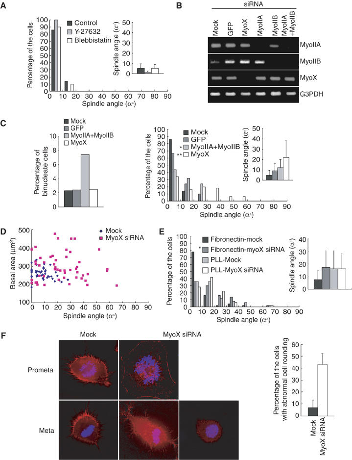
Myosin X is required for the spindle orientation parallel to the substrate surface and actin reorganization during M phase. (A) Synchronized HeLa cells on fibronectin were treated with DMSO (Control), Y-27632, or blebbistatin, and subjected to the spindle orientation analysis (histogram; n=50, inset; mean±s.d.; n=50). (B) Asynchronous HeLa-S3 cells on fibronectin were transfected with siRNAs for GFP, myosin X, myosin IIA, or myosin IIB, and incubated for 72 h. Total RNA was prepared and RNA levels of myosin IIA (first row), myosin IIB (second row), myosin X (third row), and control G3PDH (fourth row) were analyzed by RT–PCR. (C) Percentage of binucleate cells (left; n>500) and spindle orientation analysis (right; histogram; n=50, inset; mean±s.d.; n=50) in mock cells and in the cells transfected with GFP siRNA, myosin X siRNA, or myosin IIA siRNA and myosin IIB siRNA. *P>0.1 and **P<0.001, as compared with the GFP siRNA transfected cells, analyzed by F-test. (D) Spindle angles were plotted as a function of basal area in mock cells and in the cells transfected with myosin X siRNA (n=50). (E) Synchronized HeLa cells transfected with or without myosin X siRNA were plated on the coverslips coated with fibronectin or poly-L-lysine, incubated for 10 h, and subjected to the spindle orientation analysis (histogram; n=50, inset; mean±s.d.; n=50). *P<0.001, as compared with the mock, analyzed by F-test. (F) Mock cells and the cells transfected with myosin X siRNA were fixed and stained with fluorescein-phalloidine (red) and Hoechst (blue) (left). Percentage of the metaphase cells with abnormal cell rounding was measured (right).