
Figure 3.
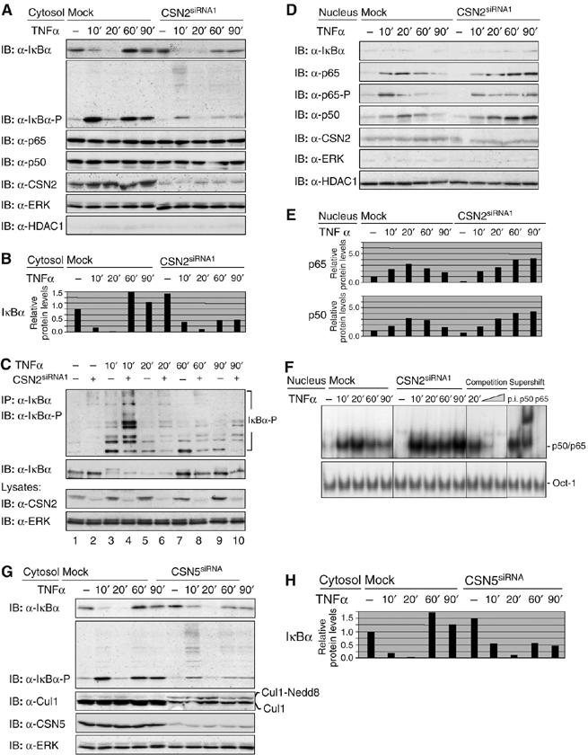
Knockdown of the CSN affects the inducible turnover of IκBα and nuclear translocation of NF-κB. Cells were transiently transfected with CSN2siRNA1 or CSN5siRNA. After 48 h, cells were stimulated for different periods of time, as indicated, with TNFα. The cells were harvested in hypotonic lysis buffer, followed by cytosolic/nuclear fractionation. Efficient knockdown of CSN2 or CSN5 (indicated panels) was analysed in cell lysates by immunodetection. In order to control the purity of cytosolic and nuclear preparations, ERK was immunodetected as a cytosolic and HDAC1 as a nuclear marker protein. (A) Cytosolic fractions of CSN2-knockdown cells were analysed by SDS–PAGE and Western blotting. To confirm TNFα-induced phosphorylation and turnover of IκBα, immunodetection of IκBα and its Ser32/Ser36-phosphorylated form was performed using specific antibodies. The p65 and p50 subunits (see the indicated panels) were immunodetected to show their predominant cytosolic localisation. (B) Quantification of the results shown for IκBα in (A) was performed as described in Figure 2B. (C) Endogenous IκBα was immunoprecipitated from cytosolic fractions of HeLa cells, transfected with CSN2siRNA1 or a nontargeting siRNA. Immunoprecipitates were analysed by SDS–PAGE and Western blotting. The IκBα protein level and its multiple high-molecular-weight forms of phosphorylated IκBα were immunodetected using IκBα- and phospho-IκBα-specific antibodies, respectively. (D) Nuclear fractions were analysed by SDS–PAGE and Western blotting. Translocated p65, Ser536-phosphorylated p65, as well as p50 (indicated panels) were immunodetected using specific antibodies. (E) Quantification of the results shown for p50 and p65 in (D). Densitometric values for protein bands were normalised to HDAC1 content in each sample and depicted relative to the values of untreated mock-transfected cells. (F) Nuclear fractions were analysed by Electrophoretic mobility shift assay (EMSA). The shifted DNA-bound NF-κB p50/p65 heterodimer and the shifted DNA-bound Oct-1 transcription factor (used as a load control) are indicated. To test the specificity, competition (using 10 and 20 ng of the nonlabelled double-stranded Igκ oligonucleotide) and antibody supershifting using anti-p50 and anti-p65 antibodies as well as preimmune serum were performed. (G) Cytosolic fractions of CSN5-knockdown cells were analysed by SDS–PAGE and Western blotting. To confirm TNFα-induced phosphorylation and turnover of IκBα, immunodetection of IκBα and its Ser32/Ser36-phosphorylated form was performed using specific antibodies. Neddylated Cul1 (Cul1-Nedd8) was immunodetected, using a Cul1-specific antibody. (H) Quantification of the results shown for IκBα in (G) was performed as described in Figure 2B.