
Figure 5.
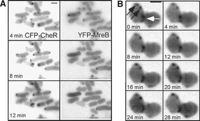
Independence of cluster positioning of the MreB filament. (A) Time-lapse image sequence showing receptor clusters (marked by CFP-CheR as in Figure 4, left panel) and MreB filaments (marked by YFP-MreB, right panel) in wild-type cells. YFP-MreB expression was induced from pDK99 with 5 μM IPTG. The cells were treated with the MreB-depolymerizing antibiotic A22 at zero time point. Disassembly of the MreB complexes did not have a visible effect on the positioning of lateral clusters. (B) Time lapse of a dividing mreB (HCB1435) cell. Constriction site (white arrow) serves as a reference point for the future polar zone. Clusters that are lateral relative to this position (black arrows) do not move within the time of the imaging, whereas the cluster at the constriction site is mobile. Clusters were marked by YFP-CheR as in Figure 1.