
Figure 4.
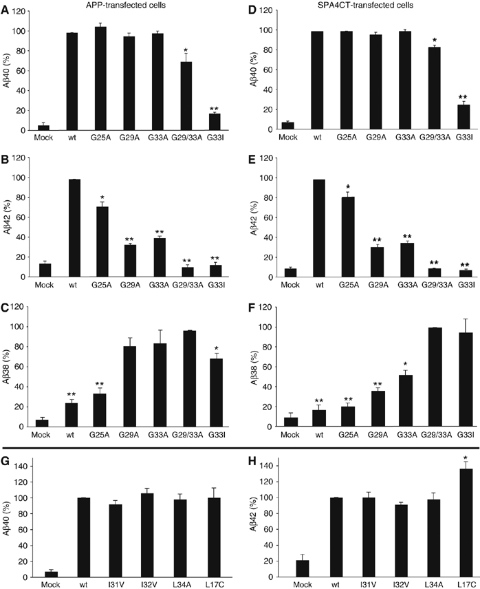
Disturbed GxxxG motifs reduce Aβ42 and increase Aβ38 levels. (A–C) Aβ40-, Aβ42- and Aβ38-specific ELISAs, respectively, with Aβ precipitated from media of stably APP-transfected SH-SY5Y cells. (D–F) Aβ40-, Aβ42- and Aβ38-specific ELISAs, respectively, with Aβ precipitated from media of stably SPA4CT-transfected SH-SY5Y cells. (A, B, D and E) The wt was set as 100% (means±s.e.m., n=3–5). Asterisks indicate significant differences to wt (*P<0.01 and **P<0.0001, Student's t-test). Single alanine mutants do not affect Aβ40 levels, but reduce the Aβ42 level significantly. The mutations G29/33A and G33I reduce Aβ40 as well as Aβ42 levels. The mutations G37A and G38A could not be analyzed by ELISA as the mutations altered epitope recognition by the monoclonal antibodies specific for Aβ40 or Aβ42. (C, F) The mutant G29/33A was set as 100% in Aβ38 ELISAs (means±s.e.m., n=3–5). Asterisks indicate significant differences to G29/33A (*P<0.01 and **P<0.0001, Student's t-test). GxxxG motif-disturbing mutations gradually increase Aβ38 levels depending on the individual glycine substitution. (G, H) Aβ40- and Aβ42-specific ELISAs from transiently or stably transfected SH-SY5Y cells. The wt was set as 100% (means±s.e.m., n=3–9). Non-glycine mutants do not affect Aβ40 or Aβ42 levels. Mutant L17C specifically increased Aβ42 levels by ∼36%.