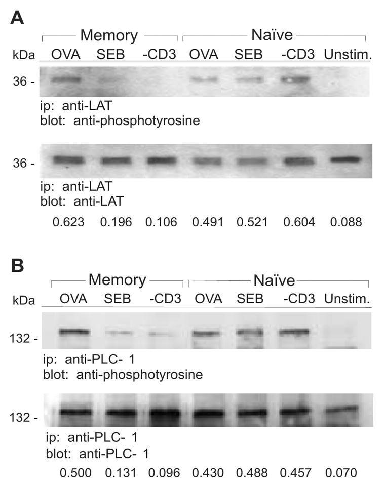
Figure 2. Decreased phosphorylation of LAT and PLC-γ1 in memory CD4 T cells stimulated with SEB and anti-CD3.

Naive and memory DO11.10 T cells were stimulated for 2 min with OVA, SEB, anti-CD3 or left unstimulated (Unstim.). A, LAT was immunoprecipitated (ip) from the T cell lysates and proteins were resolved on 12% SDS-PAGE gels followed by transfer to PVDF membranes. Immunoblotting (blot) was performed with anti-phosphotyrosine and anti-LAT Abs. B, PLC-γ1 was immunoprecipitated from the T cell lysates and proteins were resolved on 8% SDS-PAGE gels followed by transfer to PVDF membranes. Immunoblotting was performed with anti-phosphotyrosine and anti-PLC-γ1 Abs. Densitometry was performed on the immunoblots and the relative levels of phosphoprotein are expressed as a ratio relative to the total amount of precipitated protein. Data are representative of at least two independent experiments.
