
Figure 2. PKCδ overexpression in RASMCs, via adenoviral transfection, inhibits proliferation and migration.
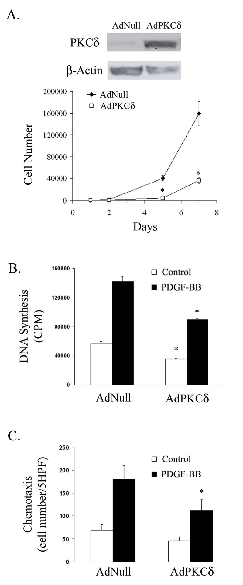
(A) RASMCs were infected with AdNull or AdPKCδ. Forty-eight hours after infection, cells were lysed and analyzed by immunoblotting. Additional infected cells were re-seeded into 24-well plates at a density of 10,000 cells per well on day 0 and maintained in 10% FBS. Cells were counted on days 1, 2, 5 or 7. (B) Following infection with AdNull or AdPKCδ, RASMCs were serum-starved for 48 h. Basal and PDGF-BB induced DNA synthesis was measured using the 3H-thymidine incorporation. (C) RASMCs were infected with AdNull or AdPKCδ. Forty-eight hours after infection, basal and PDGF-BB (5 ng/ml) induced chemotaxis was evaluated. (n=3, *p< 0.05, compared to AdNull control).