
Figure 5. PKCδ’s inhibition of migration and proliferation can be overcome by restoring ERK1/2 activity.
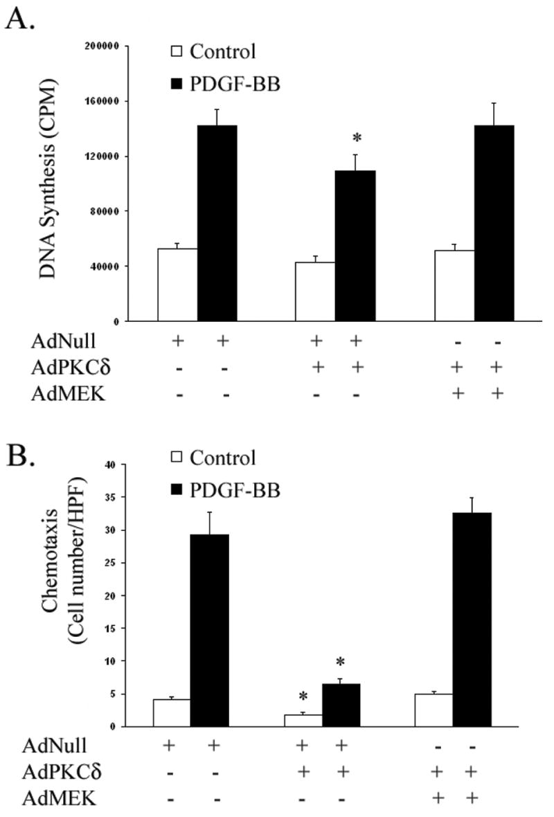
(A) Following low-serum incubation for 48 h, incorporation of 3H-thymidine was measured in A10 SMCs infected with equal quantities of AdNull, AdPKCδ, or AdMEK (60,000 total viral particles per cell) and stimulated for 24 h with or without PDGF-BB (5 ng/ml). (B) AdNull, AdPKCδ, or AdMEK infected A10 SMCs were serum-starved for 48 h. Chemotaxis with and without PDGF-BB (5 ng/ml) was evaluated. [n=3; *, p < 0.05 as compared to AdNull control]