
Figure 6.
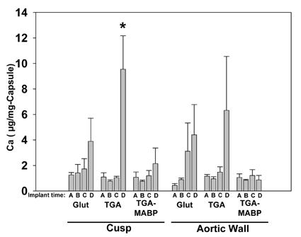
Calcium concentrations in the fibrous capsules removed from porcine aortic wall sections and porcine aortic cusp sections explanted at timepoints A= 3 days, B= 14 days, C= 21 days, and D= 90 days. Respective preparations of explants were with Glut, TGA, and TGA/MABP. Capsules showed low levels of calcification compared to the explants (see Fig. 6), and with the exception of capsules from 90-day TGA cusp explants, no statistically significant increases over time (*p<0.01).