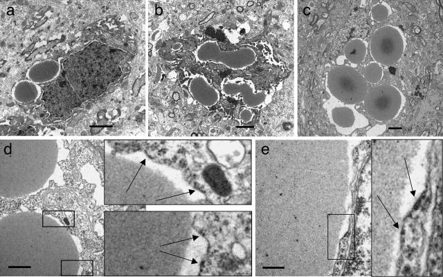
Figure 4.
Ultrastructural analysis of brains of Portland neuroserpin mice (line 793). At 6 months (a), inclusion bodies are round or oval. At 10 months (b), they are more numerous, larger, and have a more complex shape, suggesting that they may be the product of fusions. At late time points (c, 21 months), inclusions fill the entire cell, thus deforming it. In d and e, arrows indicate occasional ribosomes on the membrane surrounding inclusions in the brain of a 21-month-old Portland neuroserpin transgenic mouse. Scale bars: a–c, 2 μm; d, 1 μm; e, 0.3 μm.

