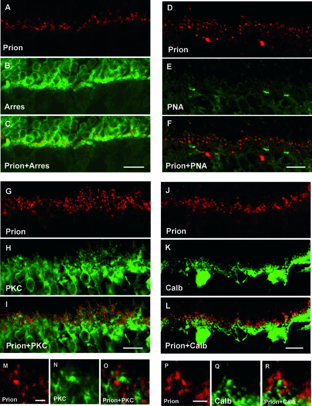
Figure 2.
Subcellular localization of the prion PrPc protein in the rat OPL. Confocal microscopic observations of rat retinal sections immunolabeled for the prion protein (red in A, C, D, F, G, I, J, L, M, O, P, R), arrestin (Arres, green in B, C), peanut agglutinin lectin (PNA, green in E, F), PKC-α, (PKC, green in H, I, N, O), calbindin (Calb, green in K, L, Q, R). The prion protein was co-localized with the arrestin labeling of the photoreceptor terminals (A–C). No prion-immunopositive structures were associated with the PNA staining of cone photoreceptor terminals (D–F). PrPc-immunopositive structures were in apposition to PKC-α-positive rod bipolar cell dendrites (G–I, M–O) and calbindin-positive horizontal cell tips (J–L; P–R). Scale bars represent 10 μm in A–L and 2 μm in M–R.

