Figure 5.
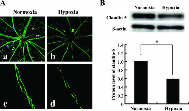
In vivo effect of tissue hypoxia on the expression of claudin-5 in retinal blood vessels. Expression of claudin-5 in retinas from mice that had been maintained under normoxia or hypoxia for 7 days was investigated by immunofluorescence (A) and Western blotting (B). The level of β-actin was used as a loading control of Western blotting. A: Claudin-5 expression in the plasma membranes of retinal blood vessels is depressed in mice under hypoxia (b, d) in contrast to the distinct expression in the mice under normoxia (a, c). The decrease in claudin-5 expression by hypoxia was distinct in the peripheral blood vessels (arrows in a) compared with the reserved expression of claudin-5 in the proximal blood vessels (arrowheads in a) (c, d). B: Tissue protein level of claudin-5 significantly decreased in the hypoxic retina compared with the normoxic retina (top panel, gel of Western blotting; bottom panel, quantitative analysis of Western blotting for the relative decrease in claudin-5 expression in hypoxic retina). *P < 0.05.

