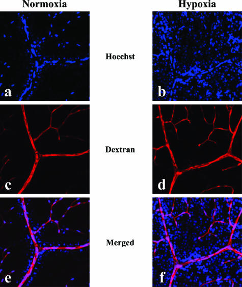
Figure 6.
Tracer experiment to evaluate the permeability of retinal blood vessels in vivo under normoxia or hypoxia. Injected tracers, Hoechst stain (a, b) and dextran (c, d), in normoxic (a, c, e) and hypoxic (b, d, f) retinas were detected under confocal microscopy. Merged views (e, f) of the signals of Hoechst stain and dextran are presented. Extensive nuclear staining by the extravasated Hoechst stain was noted in a hypoxic retina (f), whereas the stained nuclei were localized only in the vicinity of vascular lumen in a normoxic retina (e). On the other hand, no significant leakage of the injected dextran was detected in both normoxic (e) and hypoxic (f) retinas.

