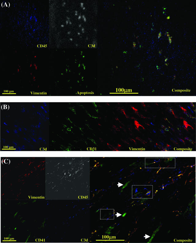
Figure 9.
Confocal laser scanning microscopy of cryostat sections of allografted heart from vimentin-immunized recipient at time of rejection. Antibody combinations are described in Materials and Methods. In A, sections stained for leukocytes (CD45-APC), C3d (Cascade Blue, pseudostained for white to represent the C3d signal), vimentin (Alexa 594), and apoptosis (terminal deoxynucleotidyl transferase dUTP nick-end labeling-FITC). The composite shows co-localization of vimentin and C3d expression on apoptosing infiltrating recipient leukocytes. In B, sections were stained for C3d (Cascade Blue), endothelial cells, CD31 (Alexa 546), and vimentin (Alexa 594). The composite shows co-localization of vimentin and C3d on endothelial cells. In C, sections were stained for vimentin (Alexa 594), leukocytes (CD45-APC, pseudostained for white to represent the CD45 signal), platelets (CD41-FITC), and C3d (Cascade Blue). The composite shows co-localization of vimentin and C3d expression on platelet (CD41+)-leukocyte (CD45+) conjugates (dotted areas). Isolated deposits of vimentin-negative unactivated platelets did not demonstrate C3d staining on their surfaces (arrowheads). Apoptosis was demonstrated by terminal deoxynucleotidyl transferase dUTP nick-end labeling staining.

