Fig. 1.

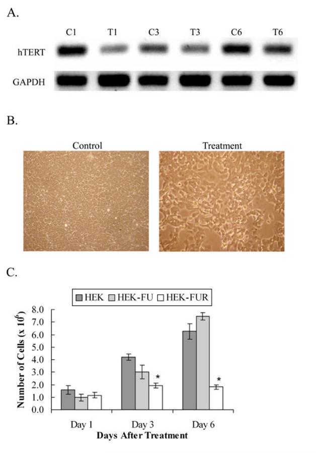
Knock down of hTERT in HEK cells from transfecting cells with dsRNA complementary to hTERT mRNA. A, PCR analysis of hTERT and GAPDH expression were performed using cDNA from treated and untreated HEK cells. C1, C3 and C6, correspond to HEK untreated control cells on treatment days 1, 3 and 6, respectively. T1, T3 and T6, correspond to HEK cells treated with 2.5 μg dsRNA complementary to a 219 bp section of hTERT mRNA on treatment days 1, 3 and 6, respectively. Levels of hTERT mRNA are reduced within 24 hours of treatment due to RNAi and the levels begin to slowly recover thereafter. GAPDH was used as a loading control. B, HEK control and dsRNA-treated HEK cells at a total magnification of 100X on day 6 after transfection. The panel with the control HEK cells shows a more densely populated culture than the treated cells. C, Cells plated at a density of 4 × 10⁵ cells and counted on days 1, 3, and 6 after dsRNA transfection. HEK: untreated control cells. HEK-FU: control cells incubated with Fugene 6 transfection reagent. HEK-FUR: dsRNA-treated cells transfected using Fugene 6 transfection reagent. Y-axis error bars represent ± standard error of the mean (SEM). * denotes statistical significance (n = 3, p < 0.05).
