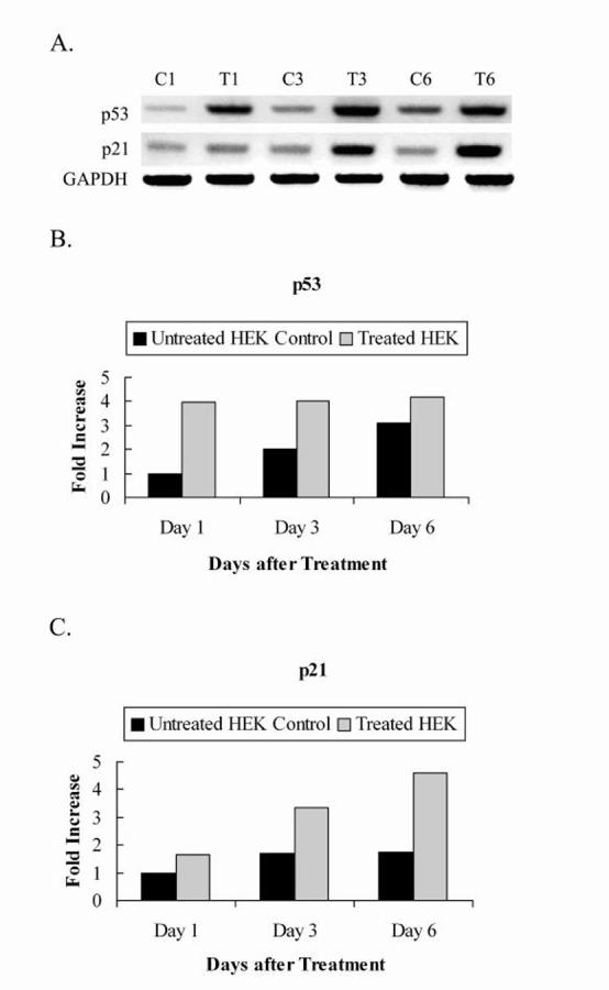
Fig. 2.

PCR analysis of p53, p21 and GAPDH expression using cDNA from HEK cells treated and untreated with synthesized dsRNA. A, 2% agarose gel electrophoresis showing p53 and p21 mRNA levels after induction of RNAi of hTERT. GAPDH was included as a loading control. Lanes C1, C3, and C6: untreated HEK control cells at days 1, 3, and 6 after treatment respectively. Lanes T1, T3, and T6: treated HEK cells at days 1, 3, and 6 after treatment respectively. B, C, Quantitations of p53 and p21 mRNA levels respectively, normalized to housekeeping gene, GAPDH. Values are from a representative gel.
