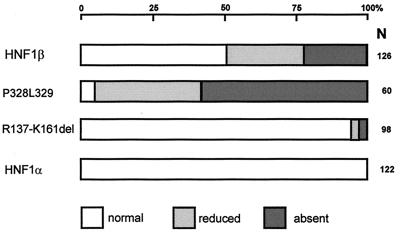
Figure 4.
Quantification of the phenotypic changes in pronephros formation observed in living Xenopus larvae. Injected normal larvae that showed green fluorescence on either the right or left side were scored at stage 45–48 for pronephros development on the injected side. The percent distribution of pronephros of normal or reduced size as well as the absence is given for the various mutants analyzed. Reduction in size of at least one-fourth was used as criteria to classify as reduced whereas in cases defined as absent, no tubular structures were visible. The number of analyzed larvae is given (N). Larvae that had unusual pigmentation, differences in the size of the eyes, or a narrower head structure were included. These minor phenotypes approximated to about 20% and were not correlated to the type of transcription factor injected.

