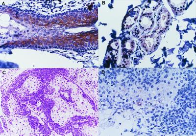
Figure 4.
Immunohistochemical detection of human Fhit in MTS tumors. (A) Fhit expression in normal hair follicle (×200); note that dense keratin horn shows nonspecific staining; (B) Fhit expression in normal sebaceous gland (×200); (C) H&E staining of an MTS case 1 sebaceous tumor; (D) Lack of Fhit expression in most cells of the case 1 sebaceous tumor.

