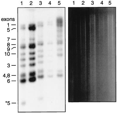
Figure 5.
Integrity of Fhit loci in murine tumors. DNA from tails and sebaceous tumors was cleaved with XbaI, electrophoresed, transferred to a membrane, and hybridized to a 32P-labeled full-length Fhit cDNA probe. Fhit exons are indicated on the left; the asterisk indicates the inactivated Fhit exon 5. Lanes 1, 3, and 4 contained DNAs from sebaceous tumors from Fhit +/− mice 21, 27, and 31; lane 2 contained DNA from the tail of Fhit +/+ mouse 25, and lane 5 contained DNA from a Swiss mouse 3T3 cell line, which exhibits a variant-sized exon 3 (obscured by another fragment) because of a polymorphism. The Fhit +/+ and +/− mice are B6129F1s, which exhibit two different alleles of exon 8. (Right) The agarose gel before blotting of the digested DNAs to the membrane; this gel illustrates that amounts of DNA loaded in individual lanes varied from ≈1 μg (lane 4) to ≈10 μg (lane 2).

