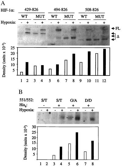
Figure 4.
Effect of internal deletions and missense mutations on HIF-1α expression. (A) Expression vectors encoding HIF-1α deletion constructs, consisting of residues 1–391 fused to 429–826, 494–826, or 508–826, either wild type (WT) or containing the S551G/T552A missense mutations (MUT), were transfected into 293 cells and exposed to 20% or 1% O2 for 4 h. Nuclear extracts were prepared, and 15-μg aliquots were subjected to immunoblot assay, which detected both endogenous full-length (FL) and recombinant deleted (Δ) HIF-1α. The expression of the deleted HIF-1α proteins was quantified by densitometry. (B) Cells were cotransfected with pSVβgal and vectors encoding HIF-1α(1–391/512–826) (lanes 1–2), His6-HIF-1α(1–391/512–826) (lanes 3–4), His6-HIF-1α(1–391/512–826/S551G/T552A) (lanes 5–6), or His6-HIF-1α(1–391/512–826/S551D/T552D) (lanes 7–8). Transfected cells were exposed to 20% or 1% O2 for 4 h, and His-tagged proteins were purified from lysates by metal-affinity resin binding. Aliquots of purified protein (normalized to β-gal expression) were subjected to immunoblot assay by using an anti-HIF-1α polyclonal Ab, and protein expression was quantified by densitometry.

