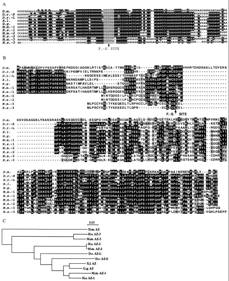
Figure 1.
Comparison between the mouse and human genes for antizyme 3 and the other known antizymes. D.m., Drosophila melanogaster; D.r., Danio rerio (S, short; L, long); X.l., Xenopus laevis; G.g., Gallus gallus; M.m., Mus musculus; H.s., Homo sapiens. (A) Comparison of the nucleotide sequences of the frameshift sites of different antizyme genes. Black background indicates nucleotide identity among at least seven antizyme genes. The frameshift (F.-S.) site is indicated with an arrow, and the UCCUGA sequence is shown with a gray background. (B) Comparison of the protein sequences of antizymes from different organisms. A black background indicates amino acid identity among at least five proteins. Gray background indicates amino acid similarity among at least six proteins. The arrow indicates the position of the frameshift site. (C) Unrooted phylogenetic tree of the antizyme proteins drawn with the clustalx program.

