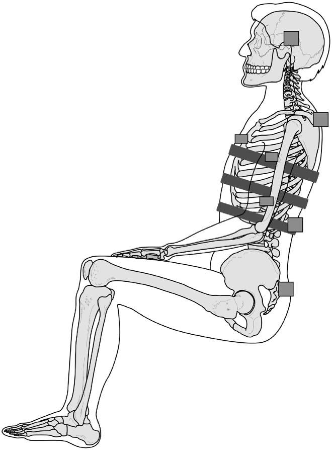
Fig. 1.

Instrumentation used in PMHS tests. Squares show accelerometers mounted to the upper and lower thoracic spinous processes, ribs and head. Three inclined rectangles show the chestbands affixed at the three levels to obtain time–deformation chest contours from which injury criteria were computed.
