Figure 4.
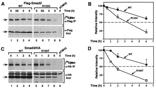
The mutation in Smad2 and Smad4 causes an increased rate of protein degradation. COS-1 cells transfected with wild-type (WT) or mutant (R133C) Flag-Smad2 (A) and wild-type (WT) or mutant (R100T) Smad4-HA (C) were labeled with [35S]methionine and then incubated in unlabeled culture medium for the indicated times. Cell lysates were subjected to immunoprecipitation, and labeled protein was visualized by autoradiography ([35S]Met). Aliquots of cell lysates were immunoblotted to determine the steady state of protein levels (α-Flag or α-HA blot). (B and D) The relative levels of Smad proteins were quantitated by laser densitometry and are expressed as the mean ± SD of three separate experiments, with the exception of the 0.5-h (B) and 1-h (D) time points, which represent single points.

