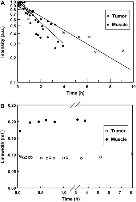
FIGURE 4.
Stability of PTM-TE in normal and tumor tissues. A small volume (20 μL) of 12 mM PTM-TE in HFB was injected directly into muscle or RIF-1 tumors grown in the hind leg of C3H mice and the intensity of the EPR signal was continuously measured using an L-band EPR spectrometer for up to 10 h. The plot (A) shows the time-course of EPR intensity data (relative to respective initial reading, displayed on a logarithmic scale) obtained from three mice per group. The decay half-life of PTM-TE was 3.3 ± 0.4 h in tumor and 2.3 ± 0.5 h in muscle. The plot (B) shows changes in the linewidth of the EPR signal during the measurement period. No significant change in the linewidth was observed.

