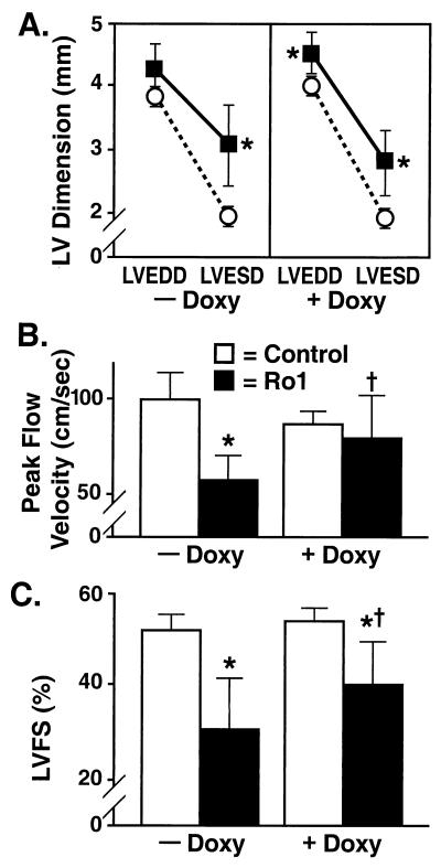
Figure 5.
Echocardiographic evidence of cardiomyopathy and recovery in mice expressing Ro1. (A) LV dimension. Mice expressing Ro1 for 8 wk showed an increase in LV end-systolic diameter (LVESD) (■, Ro1, αMHC-tTA/tetO-Ro1, n = 9; ○, control, αMHC-tTA, n = 9). After administration of doxycycline (Doxy) to these same mice for 4 wk, there was a persistent increase in LVESD and a new increase in LV end-diastolic diameter (LVEDD). (B) Peak flow velocity. After 8 wk of Ro1 expression, aortic peak flow velocity decreased by nearly 40%. After 4 wk of doxycycline administration, peak flow velocity increased significantly. (C) LVFS. After 8 wk of Ro1 expression, LVFS decreased by nearly 40%. LVFS increased significantly after suppression of Ro1 expression but remained less than LVFS in control mice. *, P < 0.05 vs. controls; **, P < 0.001 vs. controls; †, P < 0.05 vs. same mice at the previous time point.

