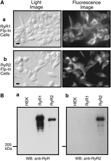
FIGURE 1.
Immunofluorescent staining and Western blot analysis of stable, inducible HEK293 cell lines expressing RyR1 or RyR2. (A) Stable, inducible HEK293 cells were fixed and permeabilized 24 h after induction by tetracycline. RyR1 or RyR2 proteins were detected using anti-RyR antibody and secondary rhodamine-conjugated antimouse IgG antibody. Light (left) and fluorescent (right) images are shown (scale bar, 10 μm). (B) Cell lysates were prepared from parental HEK293 cells (HEK), RyR1-expressing HEK293 cells (RyR1), and RyR2-expressing HEK293 cells (RyR2). RyR proteins were pulled down by GST-FKBP12.6 from the same amount of cell lysate. The GST-FKBP12.6 precipitates were immunoblotted with anti-RyR antibody (a) or anti-RyR2 antibody (b).

