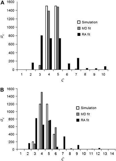
FIGURE 7.
Distribution of coefficients from MD and RA fits of (A) 4MD and (B) 8MD (see Tables 1 and 5; a Case III example). The open bars are  , the actual number of ROIs in the set with c fluorophores. The vertical bars are
  , the best-fit number of ROIs in the set with c fluorophores for the fit using MD basis histograms. The solid bars are
  , the best fit number of ROIs in the set with c fluorophores for the fit using RA basis histograms.

