
Figure 4.
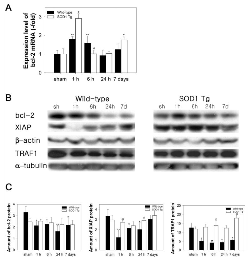
Expression levels of anti-apoptotic factors in ischemic WT and SOD1 Tg mouse brains after 30 min of tFCI. (A) QPCR from RNA obtained from damaged WT mouse brains showed an increase in bcl-2 mRNA transcription 1 h after 30 min of tFCI. In the SOD1 Tg mice, bcl-2 mRNA was up-regulated at 1 h, which is significantly higher than in the WT mice. (B) Protein levels of anti-apoptotic components in WT and SOD1 Tg ischemic mouse brains after 30 min of tFCI. Western blots showed decreased immunoreactivity of bcl-2 and XIAP in the ischemic caudate and TRAF1 in the cytosol of the WT ischemic mouse brains after tFCI. In contrast to the WT mice, increased bcl-2, XIAP, and TRAF-1 proteins were seen after tFCI in the SOD1 Tg mice. β-actin and α-tubulin were used as controls and showed no changes during the time course. sh, sham-operated mice. (C) Quantitative analysis showed that protein expression of bcl-2, XIAP, and TRAF1 decreased in the WT mice, but slightly increased in the SOD1 Tg mice (*P< 0.05, **P< 0.01, n = 4, compared with sham-operated WT mice; #P < 0.05, ##P < 0.01 comparison between WT and SOD1 Tg mice at the same time points).