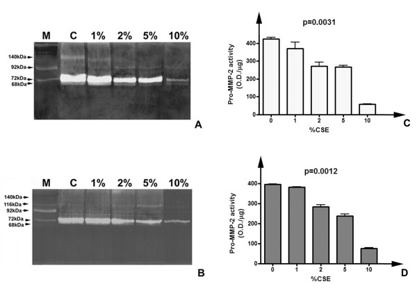
Figure 4.
Gelatinolytic pattern of HFL-1 fibroblasts conditioned media after CSE exposure. Panel of representative zymograms of conditioned media (10 μg/lane) of HFL-1 cells after 24 (A) and 48 (B) h of CSE exposure. M) Standard of human serum showing the two progelatinase bands. C) Densitometric analysis of pro-MMP-2 activity levels in conditioned media of HFL-1 cells after 24 h CSE exposure. D) Densitometric analysis pro-MMP-2 activity levels in conditioned media of HFL-1 cells after 48 h CSE exposure.

