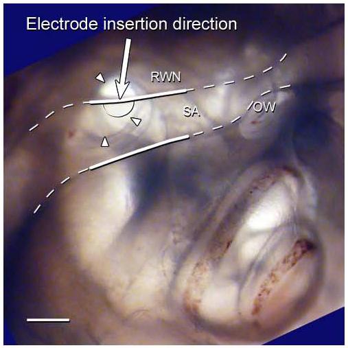
Fig. 2.

A photograph of right cochlea of a rat embedded in resin and orientated to represent the surgical view. The SA and its bony canal (white dashed line) lie partially over the RWN (arrow heads), severely restricting surgical access to the RW. The white solid lines showing the region of the SA cauterised in the surgical procedure described in this paper. The proposed cochleostomy site is shown on this figure (semi-circle solid line). The arrow represents the direction of electrode insertion. RWN – round window niche; SA – stapedial artery; OW – oval window site; Scale bar = 0.5 mm.
