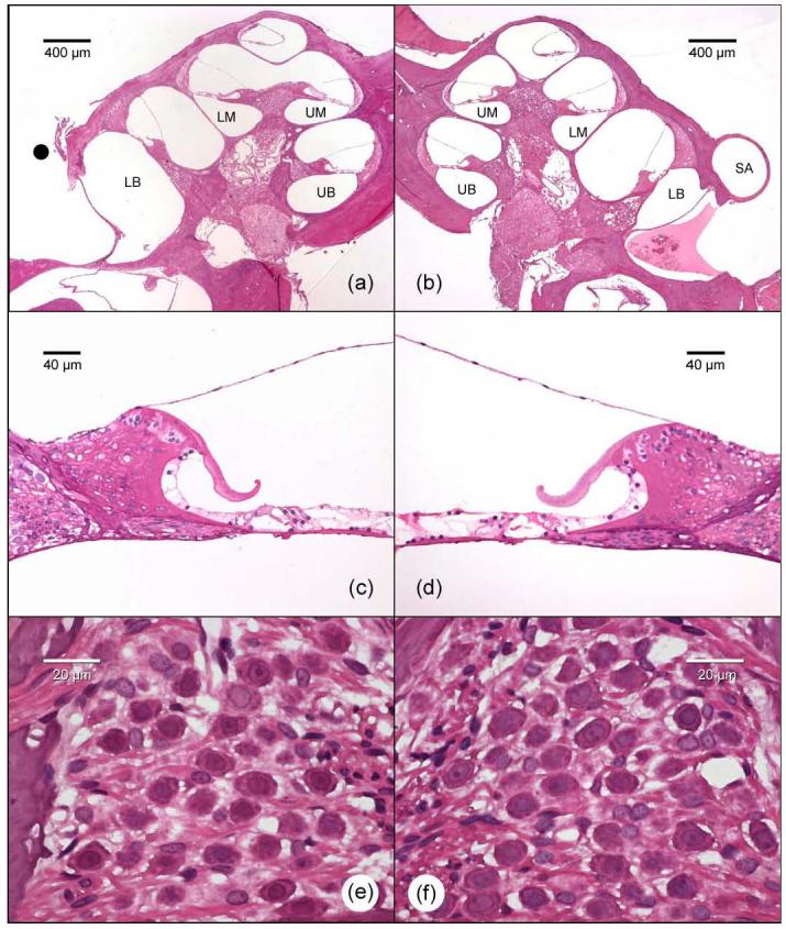
Fig. 6.

Representative low power histological images of the rat cochleae showing the normal intra-cochlear structures on a right cochlea where the SA has been cauterized (a) and its left control cochlea (b). Photomicrographs of the upper basal turns illustrating the presence of hair cells in both the SA cauterized (c) and control cochlea (d). Higher power photomicrographs illustrating well preserved SGCs in Rosenthal's canal of the upper basal turn on the SA cauterized side (e) and its contralateral control side (f). The black circle represents the site where the SA has been cauterized. LB – lower basal turn; UB – upper basal turn; LM – lower middle turn; UM – upper middle turn; SA – stapedial artery.
