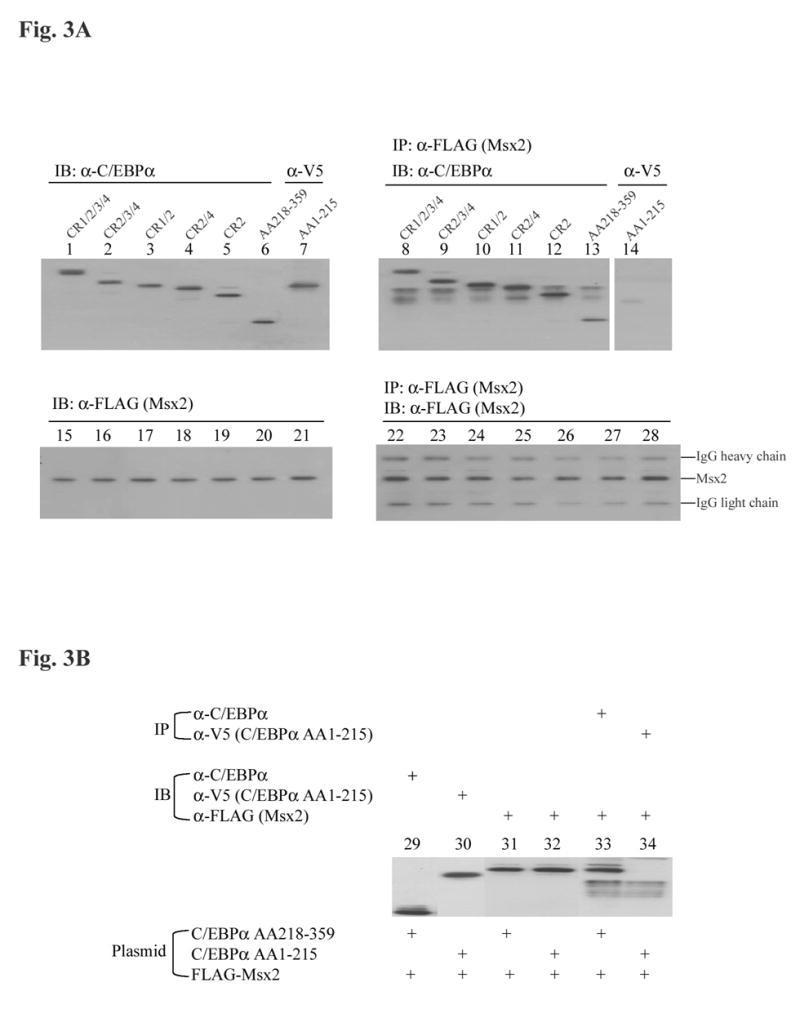
Fig. 3.

Co-immunoprecipitation of various C/EBPα truncated isoforms and Msx2 from LS8 cells. (A) In a 100-mm tissue culture dish, 2 μg of amino-terminal FLAG tagged Msx2 expression plasmid was cotransfected into LS8 cells with 2 μg of various C/EBPα truncated isoforms, CR1/2/3/4 (lane 1, 8, 15 and 22), CR2/3/4 (lane 2, 9, 16 and 23), CR1/2 (lane 3, 10, 17 and 24), CR2/4 (lane 4, 11, 18, and 25), CR2 (lane 5, 12, 19 and 26), C/EBPα AA218-359 (lane 6, 13, 20, and 27), C/EBPα AA1-215 (lane 7, 14, 21 and 28). After 24 h incubation, whole cell lysates were prepared as described under “Materials and methods”. For immunoblot (IB), 10 μg of cell lysates were electrophoresed, transferred to Immobilon-P membrane (Millipore, Billerica, MA), and immunoblotted with a C/EBPα antibody (Santa Cruz Biotechnology, Santa Cruz, CA, lane 1 to 6), a V5 antibody (Invitrogen, Carlsbad, CA, lane 7), or an anti-FLAG antibody (M2Ab, Sigma, St. Louis, MO, lane 15 to 21). 500 μg of cell lysates were subjected to immunoprecipitation (IP) with an anti-FLAG antibody (lane 8 to 14). The immunoprecipitates were then electrophoresed, transferred to Immobilon-P membrane, and immunoblotted with an anti-C/EBPα antibody (lane 8 to 13), or an anti-V5 antibody (lane 14), or an anti-FLAG antibody (lane 22 to 28). (B) Co-immunoprecipitation analysis was performed as in (A). Total cell lysates of 10 μg were immunoblotted with an antibody against C/EBPα (lane 29), V5 (lane 30), or FLAG (lane 31 and 32). Cell lysates were immunoprecipitated with an anti-C/EBPα antibody, followed by immunoblotting with an anti-FLAG antibody (lane 33); or with an anti-V5 antibody, followed by immunoblotting with an anti-FLAG antibody (lane 34).
