
Fig. 2.
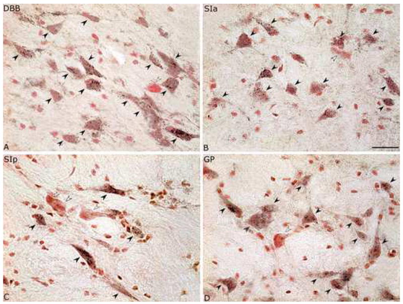
Images of PAG+/NR+ and PAG−/NR+ neurons in different BF nuclei. PAG+/NR+ cells (black arrowheads) are codistributed with PAG−/NR+ cells (white arrowheads) (on right side in same series from rat G3) in the nucleus of the diagonal band of Broca (DBB, ~A9.4, in A), the substantia innominata, pars anterior (SIa, ~A8.6 in B), the substantia innominata, pars posterior (SIp, A7.8 in C) and the globus pallidus (GP, ~A7.8 in D). Very small NR stained cells were judged to be glia and not counted as neurons. Scale bar = 25 μm.