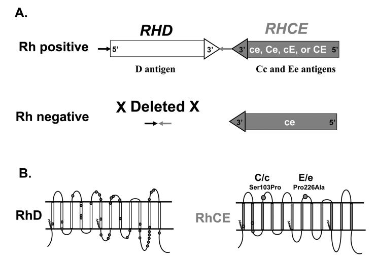
Fig. 1.
A). Diagram of the RHD and RHCE locus. The two RH genes have opposite orientation, with the 3′ends facing each other. Rh negative Caucasian individuals have a complete deletion of RHD. B). Rh proteins in the RBC membrane. The RhD and RhCE proteins are predicted to have twelve transmembrane domains. Amino acid positions that differ between RhD and RhCE are shown as dark circles on RhD. The amino acid changes responsible for C/c and E/e polymorphisms are shown on RhCE.

