
Fig. 4.
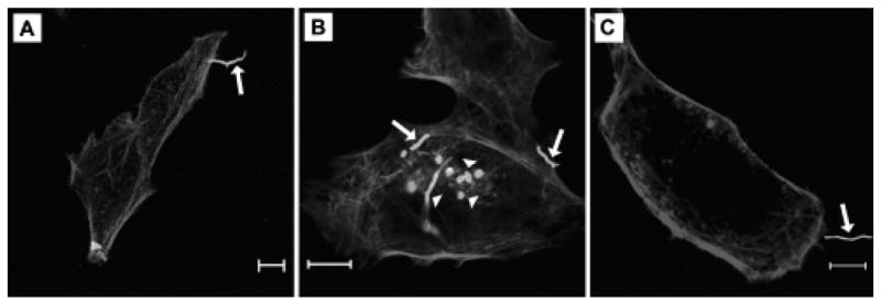
Bacterial adherence to and invasion of HeLa cells. Panels demonstrate nocardial adherence to and invasion of HeLa cells. HeLa cells were incubated with fluorescein-labeled bacteria for 2 hours and then washed and counterstained with rhodamine-phalloidin. In these panels, GUH-2 appears bright and HeLa cell actin appears dull. Panels A and B show live N. asteroides GUH-2 adhering by the filament tip and invading HeLa cells. In panel C, adherence by a heat-killed bacterium is shown. Arrows indicate bacteria adhered to cells. Arrowheads label nocardiae that appear to be within cells. The bar in each panel indicates a 10 μm distance.