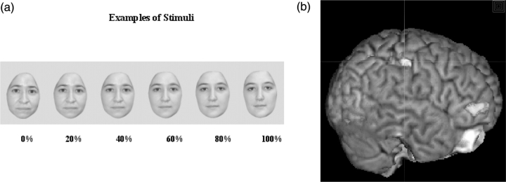
Fig. 1.
(a) Subjects viewed morphed self-images presented at random for 1 s each and used a button-box to indicate whether the image presented was ‘self’ or ‘other’. Zero% indicates no morphing (i.e. all ‘self’). (b) The right inferior parietal lobule was targeted for TMS in each individual subject by superimposing previously acquired functional imaging data at the individual subject level onto high-resolution structural images.

