Figure 2.

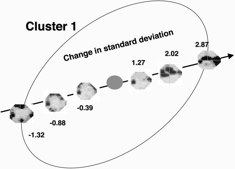
Example of fields at various SDs along axis 4 from cluster 1 in the original vB-ICA-mm space. The patterns of loss remain similar in the + and - directions with increasing severity of defect with increasing SD.

Example of fields at various SDs along axis 4 from cluster 1 in the original vB-ICA-mm space. The patterns of loss remain similar in the + and - directions with increasing severity of defect with increasing SD.